Assess

Treat
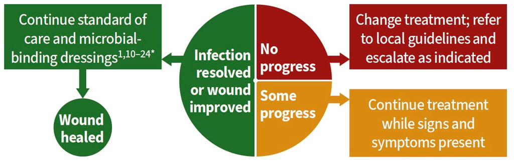
Review
Notes
*Microbial-binding dressings have a DACC coating that can control microbial burden to prevent or manage infection in a way that is not expected to contribute to antimicrobial resistance. All treatments should be used per local policy and where clinically appropriate. See below for supplementary tables and references.
Abbreviations
CEAP=Clinical, Etiological, Anatomical Pathophysiological Classification of Venous Disease; CLTI=critical limb-threatening ischaemia; DACC=dialkylcarbamoyl chloride; DVT=deep vein thrombosis; PAD=peripheral arterial disease; TIMERS=Tissue, Infection/Inflammation, Moisture balance, Edge/epithelialisation, Regeneration and repair, Social factors
Aspects of a holistic patient assessment in venous leg ulceration
– adapted from TIMERS1-6, 8-12
Patient assessment
Comorbidities
Current medication
Functionality and mobility
Nutritional assessment
Skin assessment (including skin tone)
Social factors
Surgical and medical history
Lower-leg assessment
Ankle or toe brachial pressure index
CEAP classification
Doppler/vascular ultrasound
Leg and foot pulses
Oedema
Skin perfusion
Skin temperature
Surrounding skin condition
Transcutaneous oxygen pressure
Vitals
Wound assessment
Classification
Imaging as appropriate
Location, duration, size and depth
Odour
Pain
Periwound condition
Previous investigations and treatments
Tissue biopsy (if appropriate in ≥3 months duration or atypical wound presentation)
Tissue types on wound bed (necrotic, sloughy, granulation or epithelial
Risk factors for wound infection
– adapted from the International Wound Infection Institute1, 20, 21
Patient risk factors
Alcohol, smoking or illicit drug use
Conditions associated with hypoxia or poor perfusion (e.g. anaemia, cardiac disease, respiratory disease, peripheral arterial disease, renal impairment or rheumatoid arthritis)
Connective tissue disorders (e.g. Ehlers-Danlos syndrome)
Corticosteroid use
Immune disorders (e.g. acquired immune deficiency syndrome
Lymphoedema
Malnutrition or obesity
Neuroarthropathy
Peripheral arterial disease (inc. ischaemia)
Peripheral neuropathy (sensory, motor and autonomic)
Poor adherence to treatment plan
Poorly controlled diabetes
Radiation therapy or chemotherapy
Wound risk factors
Atypical wounds
Duration of wound
Foreign body presence (e.g. drains, sutures or wound dressing fragments)
Haematoma
Impaired tissue perfusion
Increased exudate and oedema that is not adequately managed
Involvement of tissue deeper than skin and subcutaneous tissues (e.g. tendon, muscle, joint or bone)
Necrotic or sloughy wound tissue
Environmental risk factors
Hospitalisation (due to increased risk of exposure to antimicrobial-resistant microorganisms)
Inadequate hand hygiene and aseptic technique
Inadequate management of moisture (e.g. due to exudate, incontinence or perspiration)
Unhygienic environment (e.g. dust, unclean surfaces, or presence of mould/mildew)
Signs of sepsis⁶
Sepsis is a life-threatening condition in which the body's response to infection causes injury to its tissues and organs. Organ dysfunction is a key component in any diagnosis of sepsis.
Act on any of the following red flags:
S. Slurred speech or confusion
E. Extreme shivering or muscle pain
P. Passing no urine (in a day)
S. Severe breathlessness
I. It feels like you are going to die
S. Skin mottled or discoloured

Guidance


DACC™-coated dressings instructions for use

click for REFERENCES
